
Animal Inside Out at the NHM with Body Worlds- 04.03.12
The latest dose of natural inspiration from our resident zoologist and London-based editor, Justine Aw.
As soon as we heard about a new exhibition that would bring Body Worlds to animals, I knew we had to see what the fuss was all about. This morning we had our chance to check out Animal Inside Out, the awesome new temporary exhibit at the Waterhouse Gallery of the Natural History Museum in London that gives visitors a look beneath the skin of some of nature’s most spectacular animals. The exhibition is adapted from Gunther von Hagens’ Body Worlds and jointly presented by Body Worlds and the Natural History Museum. More photos of the exhibit and more details on how plastination works on the next page!
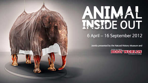
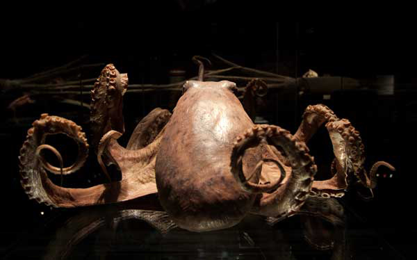
Set to open to the public this Friday, April 6th, the brand new exhibit showcases nearly 100 plastinated animals and capillary specimens and will run until the 16th of September. As in Body Worlds, the incredible specimens offer an insight into the inner workings of the animals, bringing anatomy to the fore and offering a unique perspective on these species. The techniques from Body Worlds are applied to the animals, with many preserved by plastination, a process by which water and fats are extracted and replaced with polymers in a vacuum. To create the capillary specimens, polymers are injected into the circulatory system, then the rest of the tissues dissolved using various chemicals.
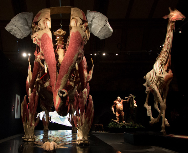
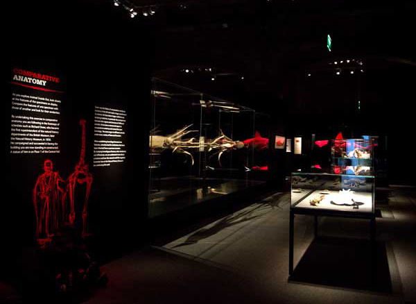
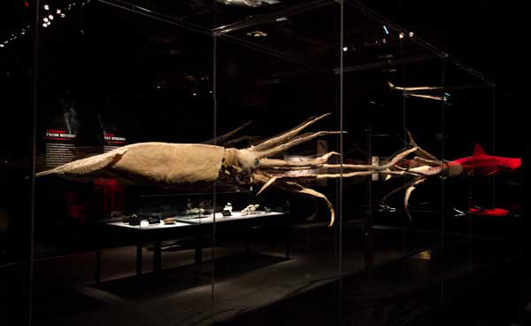
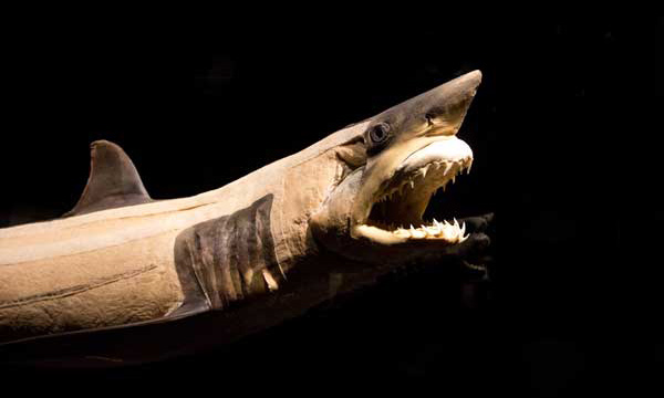
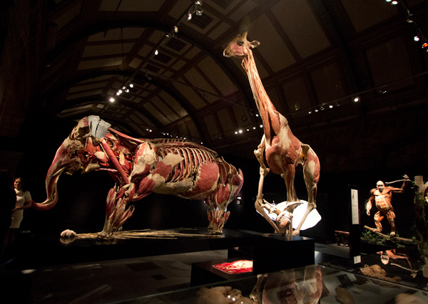
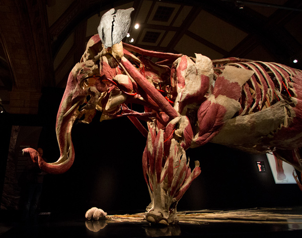
The results are phenomenal and there are plenty of oohs and ahhhhs around every corner as visitors see inside a wide variety of body plans. The exhibition combines traditional anatomical displays such as skeletal mounts with everything from slices through fishes to staggering plastinated elephants and capillary sharks. The specimens really capture the intricacy of physiology, though it is rather overwhelming and doesn’t shy away from being a bit macabre or even grotesque!
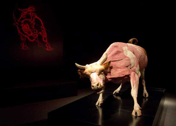
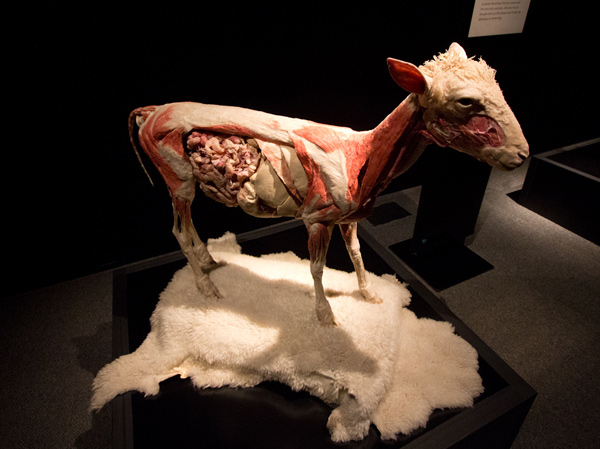
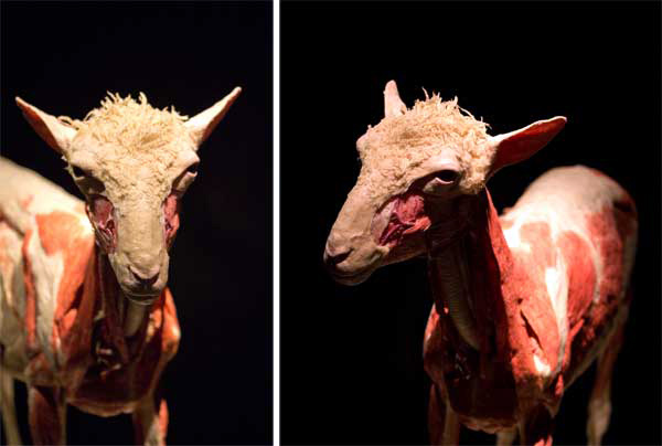
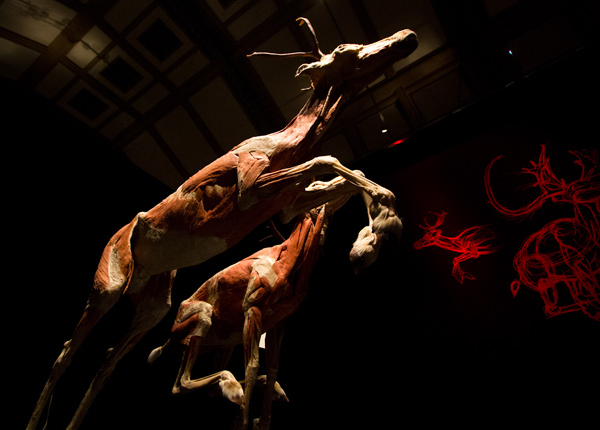
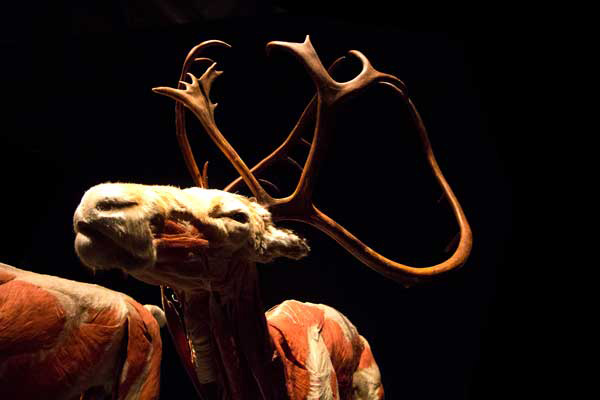
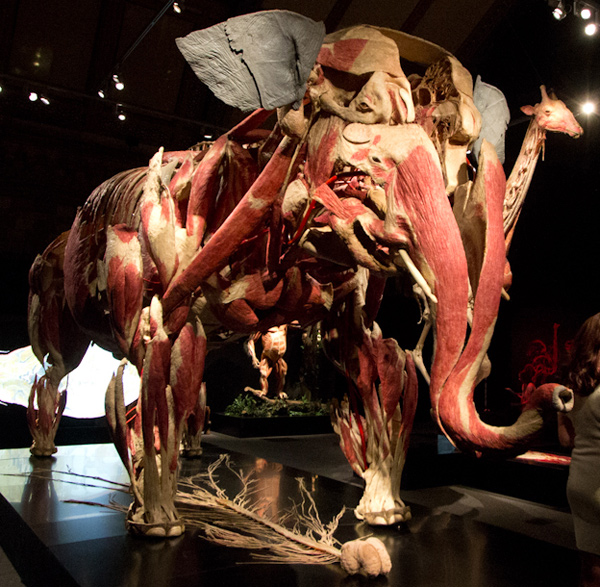
As in the human version of Body Worlds, the bodies are arranged in realistic action poses, allowing visitors to see how muscles and tendons work to make those actions possible.
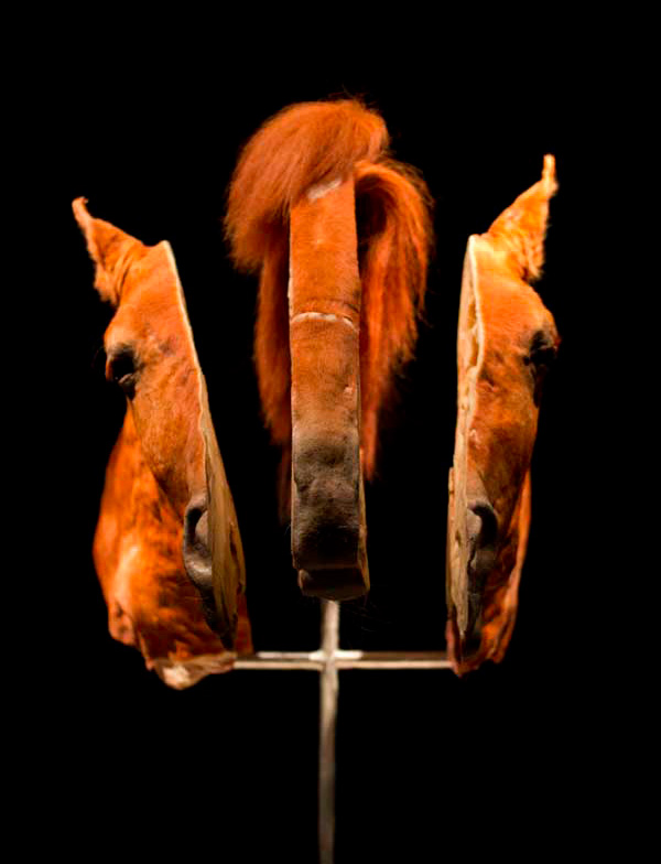
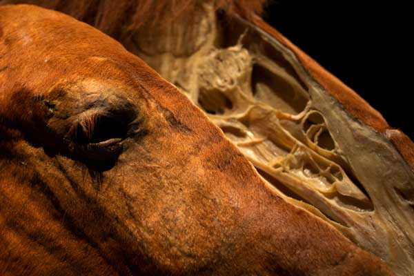
Despite being neither the largest or grandest, this was one of my favorite specimens, the trisected horse’s head that offers tantalizing glimpses inside and looks stunning from just about every angle.

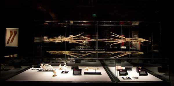
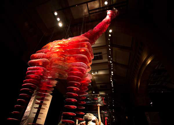
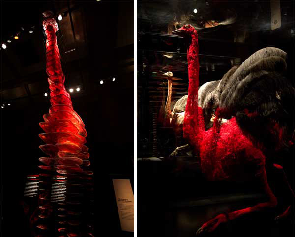
The presentation of this selection of dorsal slices through the giraffe is pretty staggering, especially when seen next to the plastinated giraffe!
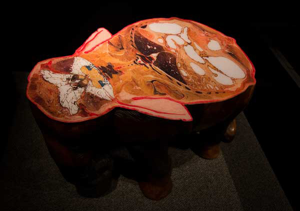
Though overshdowed by the enormous elephant specimen, I loved this little elephant model that shows visitors what a dorsal slice through the animal looks like.
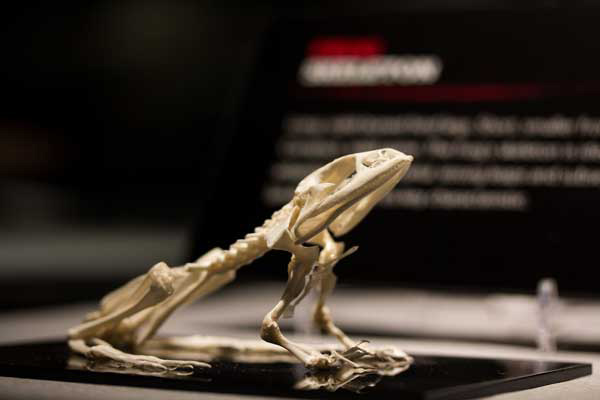
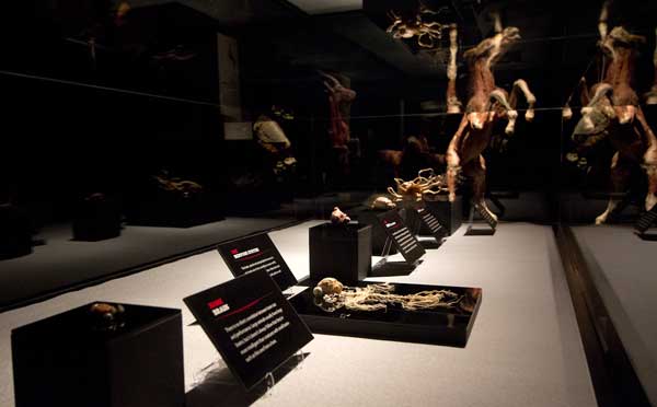
The exhibit also includes some more classical techniques such as skeletal mounts, like this little frog, and nervous system dissections which complement the new techniques.
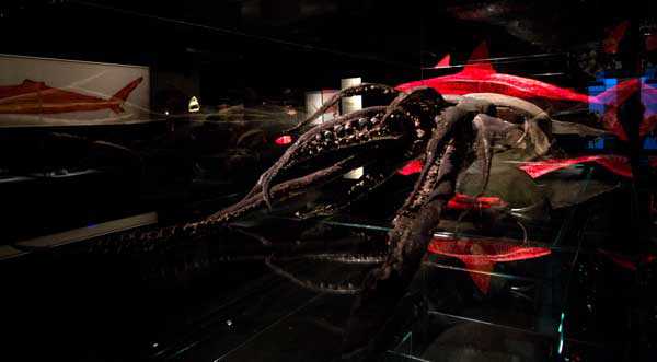
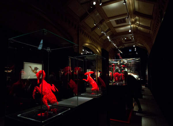
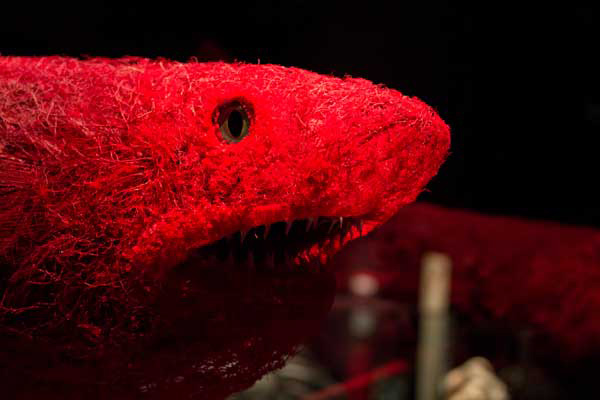
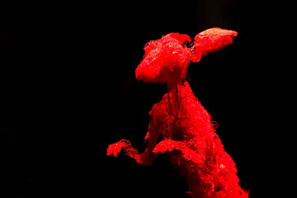
Stunning capillary specimens, created by injecting a polymer into the circulatory system, then dissolving away tissues to leave an impressive meshlike network of capillaries that perfectly capture the form of the animal, like these clearly recognizable duck, shark and rabbit.
Getting up close to these enormous specimens really reminds you of how big they are! But the comparative approach developed through the exhibition also draws so many parallels to their smaller domestic relatives.
And for those interested in the process, the Institute for Plastination team take the following steps to plastinate an animal:
Embalming and dissection First the animal has formalin pumped into its body to stop the natural process of decay and to kill any bacteria. Then it is decided what particular structure the animal is going to show and skin, fatty and connective tissues are removed as required.
Removal of body fat and water The animal specimen is placed into an acetone bath. Acetone is a solvent that replaces the water and fatty tissues that remain in the body.
Forced impregnation After this, the specimen is immersed in a liquid polymer and placed in a vacuum chamber. The vacuum sucks the acetone out of the animal’s tissues as it evaporates and the resulting vacuum in the specimen forces the polymer solution to enter in its place. This process of removing the acetone and forcing in the polymer solution continues until all of the animal’s tissues have been totally saturated.
Positioning While the specimen is still malleable it is placed into its final position for display and then held in place until it sets using wires, clamps and needles.
Curing Finally the specimen is hardened using gas, light or heat depending on the material used. This ensures the specimen lasts for a long time and can be used in exhibitions and for education.


This is such an awesome exhibition, I’m going to London on the 6th of April so I’m definitely going to check it out!
----- Shane 04.04.12 08:45